
Figure 1.
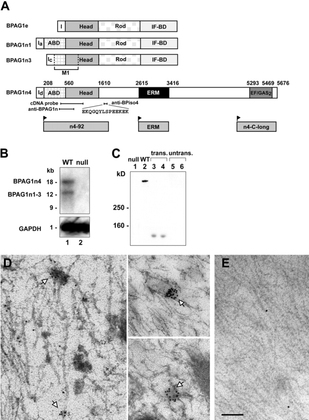
Characterization of BPAG1n4. (A) Schematic of the domain structures of BPAG1 isoforms. I, first exon of each isoform; BPAG1e, the epithelial isoform; ABD, actin-binding domain; IF-BD, intermediate filament-binding domain; M1, microtubule-binding domain. Amino acid residue numbers denote functional domain boundaries in BPAG1n4. Individual domains and regions used in this work are illustrated below. Anti-BPAG1n recognizes all BPAG1 isoforms. (B) Northern blot analysis of mouse dorsal root ganglion (DRG) RNA. (probes) A 1.6-kb cDNA head domain fragment (top; 4-d exposure); GAPDH as internal control (bottom; 1-h exposure). (C) Protein expression of BPAG1n4 in mouse brain. Blot was probed with anti-BPiso4. COS-7 (lane 3) and NIH 3T3 cells (lane 4) were transfected with an expression construct for n4-92 (A, bottom) encompassing the epitope for anti-BPiso4. (untrans.) Untransfected COS-7 (lane 5) and NIH cells (lane 6). (D–E) ImmunoEM reveals subcellular localization of BPAG1n4 in dorsal roots. (D) BPAG1n4 labeling with anti-BPiso4 was visualized with 12-nm gold particles conjugated with secondary antibody. White arrows indicate BPAG1n4 on vesicle-like structures in association with microtubules. (E) Negative control, secondary antibodies alone. Bar: (D) 300 nm; (E) 400 nm.