Figure 1.
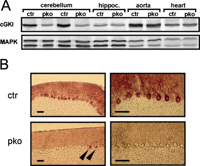
Conditional ablation of cGKI in cerebellar PCs. (A) Western blot analysis of cGKI expression (top) in various tissues of control mice (ctr) and cGKIpko mice (pko). Equal loading of protein extracts from tissues of control and cGKIpko mice was confirmed by staining the blot with an antibody against p44/42 MAPK (bottom). (B) Immunohistochemical detection of cGKI on sagital cerebellar sections of control mice (ctr, top) and cGKIpko mice (pko, bottom). Arrowheads indicate cGKI-positive PCs in cGKIpko mice. Bars, 50 μm.

