
Figure 1.
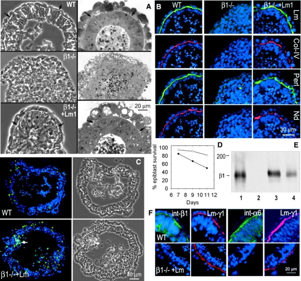
Laminin induction of basement membrane and epiblast in β1-integrin–null EBs. Wild-type and β1-integrin–null ES cells were grown in suspension for 7 d, the latter maintained alone or in the presence of laminin-1 (25 μg/ml). (A) Phase micrographs (left) and methylene blue-stained sections (right) of wild-type (top), untreated (middle) and laminin-1–treated (bottom) β1-integrin–null EBs. (B) Immunofluorescence micrographs of consecutive sections of the above EBs stained with DAPI (nuclei, blue) and antibody to laminin-γ1 (Lm), type IV collagen (Col-IV), perlecan (Perl) and nidogen (Nd). (C and D) β1-integrin–null EBs were treated with 25 μg/ml laminin-1 and cultured for 7–11 d. TUNEL (green) and DAPI (blue) costaining revealed developing segmental (arrow) apoptosis (plot was calculated after subtracting EBs with full-thickness segmental apoptosis from the total). (E) Immunoblot detection of the β1-integrin subunit in wild-type (lane 1), β1-integrin–null (lane 2), dystroglycan-null (lane 3), and γ1-laminin–null (lane 4) EBs. (F) Immunofluorescence micrographs showing β1-integrin (first frame), α6-integrin (third frame), and γ1-laminin (second and fourth frames) of wild-type (top) and laminin-treated β1-integrin–null EBs (bottom).