
Figure 5.
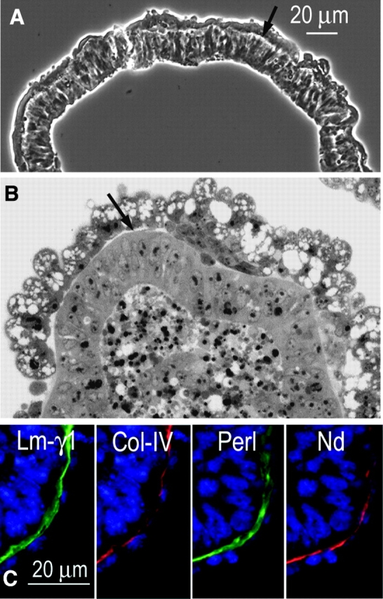
Basement membranes assemble in dystroglycan-null EBs. Dystroglycan-null ES cells, suspended at the first passage from feeder cell layers, were allowed to form EBs for 5 d in the absence of any treatment. (A) Phase micrograph and (B) methylene blue–stained section show epiblast differentiation, cavitation, and thin basement membranelike structures (arrows). (C) EBs, visualized by immunofluorescence, reveal a subendodermal basement membrane pattern costaining with antibodies for γ1-laminin, type IV collagen, perlecan, and nidogen.