
Figure 7.
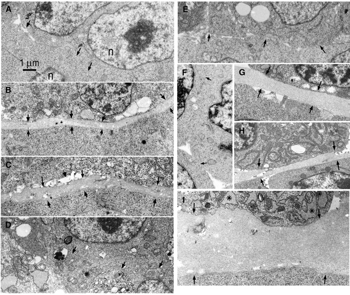
Ultrastructure of EBs. Wild-type (R1), γ1-laminin–null, β1-integrin–null, and dystroglycan-null EBs, untreated and laminin-treated, were examined by electron microscopy after incubation in suspension culture for 5–7 d. The regions containing junctions of the endodermal layer and ICM or epiblast layer are shown. (A) β1-Integrin–null embryoid body, 7 d. Endoderm (above arrows) was present; however, neither basement membrane nor epiblast differentiation is present. Arrows indicate endodermal/ICM cell boundary and n indicates nucleus. (B) Wild-type embryoid body (7 d) reveals basement membrane between endoderm and epiblast (between arrows). (C) β1-Integrin–null embryoid body treated with laminin-1 (25 μg/ml, 7 d). Basement membrane (arrows) is located between endoderm and epiblast layers. Scattered small clefts (arrowhead) located between cell and matrix were present more frequently in these EBs compared with wild-type. (D) γ1-Laminin–null EB, 7 d. No basement membrane was detected at the endoderm/ICM cell boundary (arrows). (E) γ1-Laminin–null EB treated with laminin-5 (7 d). Endodermal differentiation in the absence of basement membrane was seen. Endodermal/ICM interface indicated by arrows. (F) γ1-Laminin–null EBs treated with nonpolymerizing laminin-1 (25 μg/ml, 7 d). No basement membrane or epiblast differentiation was detected. (G) γ1-Laminin–null EBs treated with (polymerizing) laminin-1 (25 μg/ml, 7 d). Note prominent basement membrane between endoderm and epiblast layers (arrows). (H and I) Dystroglycan-null EBs, 5 d. Note typical basement membrane (H) lying between endoderm and epiblast cell layers. The RER (asterisk) of the endoderm is dilated.