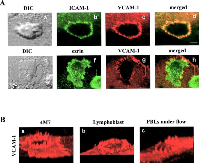
Figure 6.
Formation of the endothelial docking structure for adhered lymphocytes under flow. (A) Transendothelial migration assay of peripheral blood lymphocytes under fluid shear conditions. After 10 min of perfusion, cells were fixed and stained for ICAM-1 (b and d, green), VCAM-1 (c, d, g, and h, red), and ezrin (f and h, green). The corresponding DIC images are shown in panels a and e. Merged images are shown in panels d and h. Bar, 3.5 μm. (B) Three-dimensional reconstruction of VCAM-1 staining during 4M7 cell (a), T lymphoblast (b), or PBL under flow (c) adhesion.

