
Figure 1.
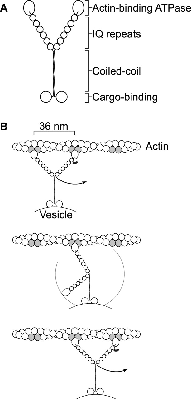
Structure and model of myosin-V. (A) Schematic diagram of myosin-V (Cheney et al., 1993). NH2-terminal actin-binding ATPase domain sequences, the six IQ repeats, the coiled-coil region, and COOH-terminal globular domain sequences are conserved in animals, fungi, plants, and slime mold. (B) Swinging lever arm model proposed for the walking of a myosin-V dimer along actin (Rief et al., 2000; Mehta, 2001). Conformational changes in the actin-binding domain during its ATPase cycle (thick arrow) rotates the lever arm, resulting in movement (thin arrow). The repeated binding sites are highlighted in gray.