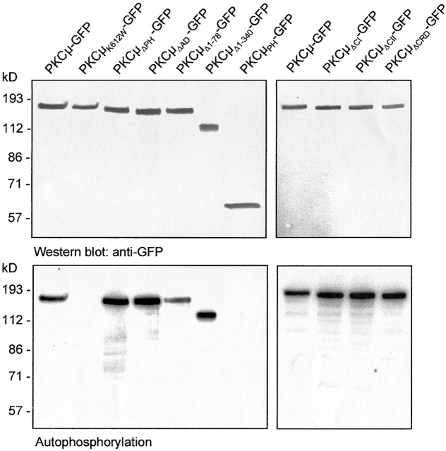
Figure 2.
Expression and in vitro phosphorylation of PKCμ-GFP fusion proteins. HEK293 cells were transfected with the indicated constructs. 40 h after transfection cells were lysed and PKCμ-GFP was immunoprecipitated using an anti-GFP antibody and subjected to Western blotting (top) and in vitro autophosphorylation (bottom). Shown are autoradiographs after overnight exposition.

