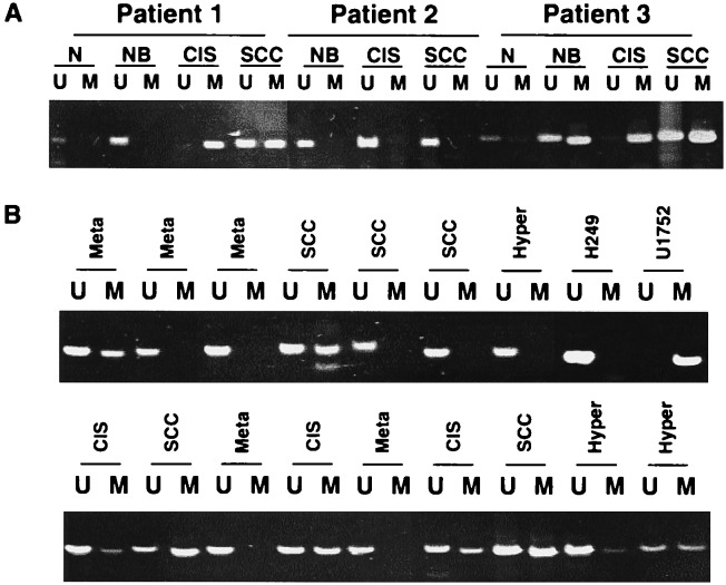
Figure 3.
Methylation of p16 in premalignant lesions, CIS, and SCC. (A) Lymphocytes microdissected from in and around the SCC constituted the normal (N) tissue for analysis. Other areas analyzed included normal appearing bronchus (NB) within the cancer field, adjacent CIS, and SCC obtained from three patients. (B) Precursor lesions and SCCs obtained through biopsy or lobectomy from several different cases. Lesions microdissected included basal cell hyperplasia (Hyper), squamous metaplasia (Meta), CIS, and SCC. Cell lines H249 and U172 serve as positive controls for detecting unmethylated (U) and methylated (M) p16 alleles, respectively by MSP. A PCR product of the appropriate molecular weight (151 bp for U, 150 bp for M) indicates the presence of unmethylated and/or methylated p16 alleles in that sample.

