
Figure 8.
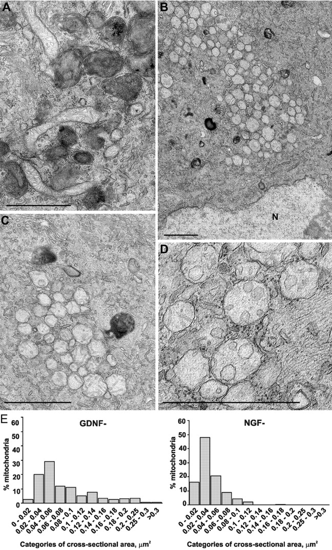
Mitochondria of NGF-deprived, but not GDNF-deprived, sympathetic neurons are structurally changed. (A) Typical view of a GDNF-deprived neuron with numerous dark autophagic profiles and several nonclustered elongated mitochondria with normal cristae. (B) An NGF-deprived neuron with several dark autolysosomes and large number of round clustered mitochondria with changed cristae. N, nucleus. (C) Higher magnification of the mitochondrial cluster with vesicular cristae and one membrane in an NGF-deprived neuron. (D) Mitochondria whose cristae and inner membrane are altered to a different extent in an NGF-deprived neuron. (E) Distribution of mitochondrial profiles from the sections of GDNF-deprived (n = 201) and NGF-deprived (n = 317) neurons according to their cross-sectional areas. Size categories are shown as a percentage of all mitochondrial profiles. Bars, 1 μm.