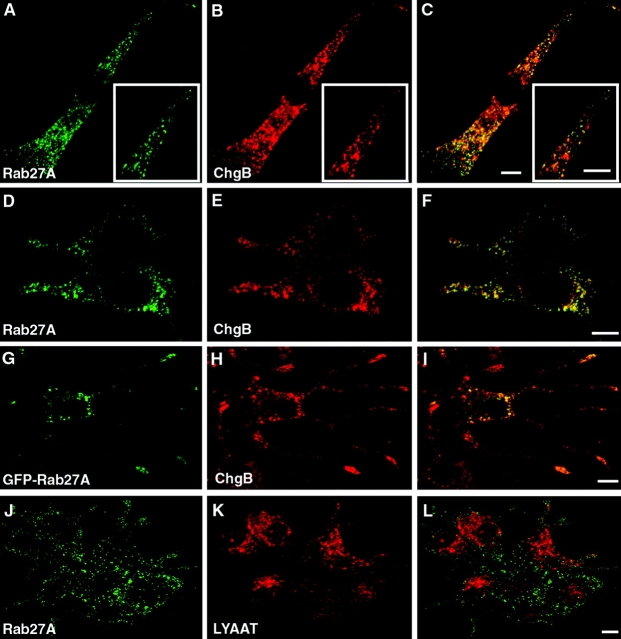
Figure 1.
Localization of Rab27A on SGs by confocal immunofluorescence. Chromaffin cells (A–C) were double imaged for endogenous Rab27A (A) and chromogranin A/B (B). Discrete punctate structures were observed in A and B; most of them precisely coincided. See the overlaid image (C) and the detail shown at higher magnification (inset). Anti-Rab27A (D) and anti–chromogranin A/B (E) also stained discrete structures that coincided (F, overlay) in PC12 cells. GFP–Rab27A (G) transiently expressed in NGF-treated PC12 cells also colocalized with chromogranin B–positive structures (H) (I, overlaid image). Note the enrichment of Rab27A and chromogranin B at the tip of neurites. In contrast, Rab27A (J) did not colocalize with LYAAT, a lysosomal marker (K and L). Bars, 5 μm.

