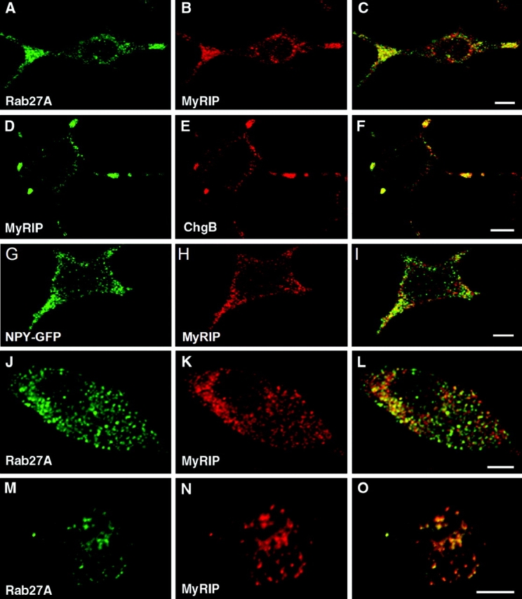
Figure 2.

Confocal immunofluorescence localization of MyRIP on SGs. PC12 cells (A–I) were double imaged for endogenous Rab27A (A) and MyRIP (B) showing discrete punctate structures; most of these structures coincided (C, overlaid image), especially in the neurites. PC12 cells were transfected with pCMV-MyRIP and double imaged for MyRIP (D and H) and chromogranin B (E) or NPY–GFP (G); overlaid images indicate that MyRIP colocalized partly with chromogranin B (F) and NPY–GFP (I). Chromaffin cells (J–O) were imaged for endogenous Rab27A (J and M) and MyRIP (K and N). Many structures were double labeled, as indicated by the overlaid image of a cell sectioned close to the apex (O). Bars, 5 μm.
