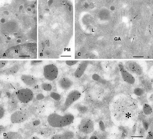
Figure 3.

Ultrastructural localization of Rab27A in PC12 and chromaffin cells. Ultrathin cryosections of PC12 (A–C) and chromaffin cells (D) were single or double immunogold labeled for Rab27A (10-nm gold particles, A–C) or Rab27A (15-nm gold particles, D) and chromogranin A/B (10-nm gold particles, D). (A and B) Specific labeling of dense core granules of PC12 cells with anti-Rab27A antibodies. A restricted number of dense granules show high density of labeling for Rab27A (arrows), whereas others are negative. (C) Rab27A-positive tubulo-vesicular structures (arrows) are often observed in the Golgi region. (D) In chromaffin cells, Rab27A localizes to chromogranin-positive SGs (arrows) and to immature granules (star). N, nucleus; PM, plasma membrane; GA, Golgi apparatus. Bars, 200 nm.
