
Figure 1.
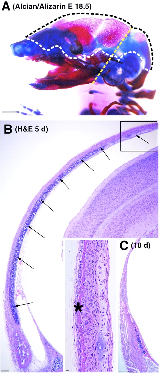
Morphology of cartilage anlagen and membranous bones in the wild-type mouse calvarium. (A) Gross morphology of the skull of an E18.5-d-old mouse embryo stained with alizarin red/alcian blue. The dotted line marks the region of membranous ossification. Note the extensive cartilaginous anlagen in the region where parietal and interparietal bones develop. The yellow dotted line indicates the plane of sectioning used in preparing histological material, as shown in B. (B) Section through the skull of a 5-d-old mouse, demonstrating the parietal cartilage (arrows) and the leading edge of the developing parietal bone (inset, asterisk). The inset shows a higher magnification of the area boxed in B. Note that parietal bone grows outside of the parietal cartilage rudiment. The cartilage matrix is not stained by hematoxylin (has lost the basophilia that indicates a normal proteoglycan content) in the region where membranous ossification is in progress. (C) Low-power magnification of parietal bone from 10-d-old wild-type mouse demonstrating complete removal of the parietal cartilage. Bars: (A) 1 mm; (B) 100 μm; (B, inset) 10 μm; (C) 333 μm.