Figure 6.
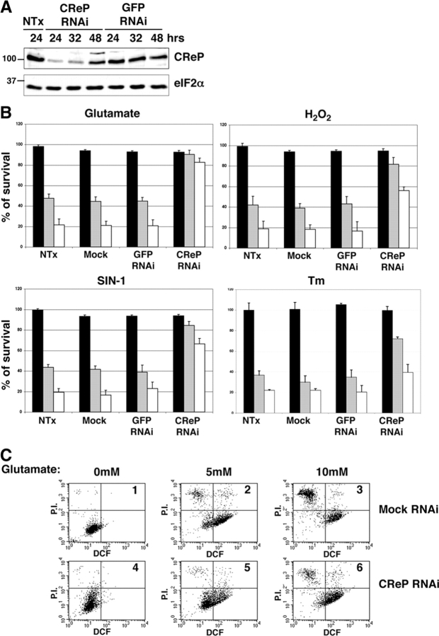
CReP RNAi promotes a stress-resistant state. (A) Immunoblot of endogenous CReP in lysates of nontransfected HT22 cells and cells transfected with a small interfering RNAi directed to CReP or to GFP. Lysates were harvested for immunoblot at the indicated time. (B) Survival, measured by MTT assay after exposure to glutamate (7 h at 5 mM, gray bar; or 10 mM, white bar), H2O2 (7 h at 1 mM, gray bar; 2 mM, white bar), SIN-1 (7 h at 1.5 mM, gray bar; 3 mM, white bar) and tunicamycin (12 h at 0.125 μg/ml, gray bar; 0.25 μg/ml, white bar followed by 24 h recovery). Cells were nontransfected, mock transfected with no siRNA, or transfected with siRNA to GFP (a control) or CReP. Shown are mean ± SEM of experiments performed in triplicates and reproduced three times. The MTT signal of the untreated, nontransfected cells is arbitrarily set at 100. (C) Dual channel FACscans of dichlorofluorescein fluorescence (DCF axis, reporting on endogenous peroxides) and propidium iodide fluorescence (P.I. axis, reporting on permeabilized, damaged cells) of untreated (boxes 1 and 4) and glutamate-treated (boxes 2, 3, 5, and 6) mock-transfected (boxes 1–3), and CReP RNAi-transfected (boxes 4–6) HT22 cells. The fraction of cells in each quadrant of the FACS® is indicated in Table I.

