Figure 2.
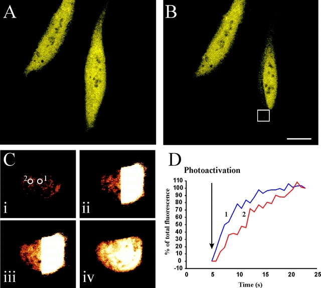
Demonstration that hippocalcin–EYFP is freely diffusible in resting cells. (A) HeLa cells were transfected to express hippocalcin–EYFP, which showed a diffuse cytosolic and nuclear localization. (B) One cell was locally photobleached within the indicated box for 30 s leading to a loss of cytosolic fluo-rescence. Nuclear fluorescence in the neighboring cells and in the nucleus of the photobleached cell was unaffected. (C) A cell transfected with pHippo-PA-GFP was illuminated with high-intensity 430-nm light on its right hand side to photoactivate the hippocalcin-PA-GFP. The images i–iv are shown before activation (i) and 2 (ii), 5 (iii), and 15 (iv) s after activation. (C) Time course of GFP fluorescence increases measured in the indicated regions of interest. Bar, 10 μm.

