
Figure 8.
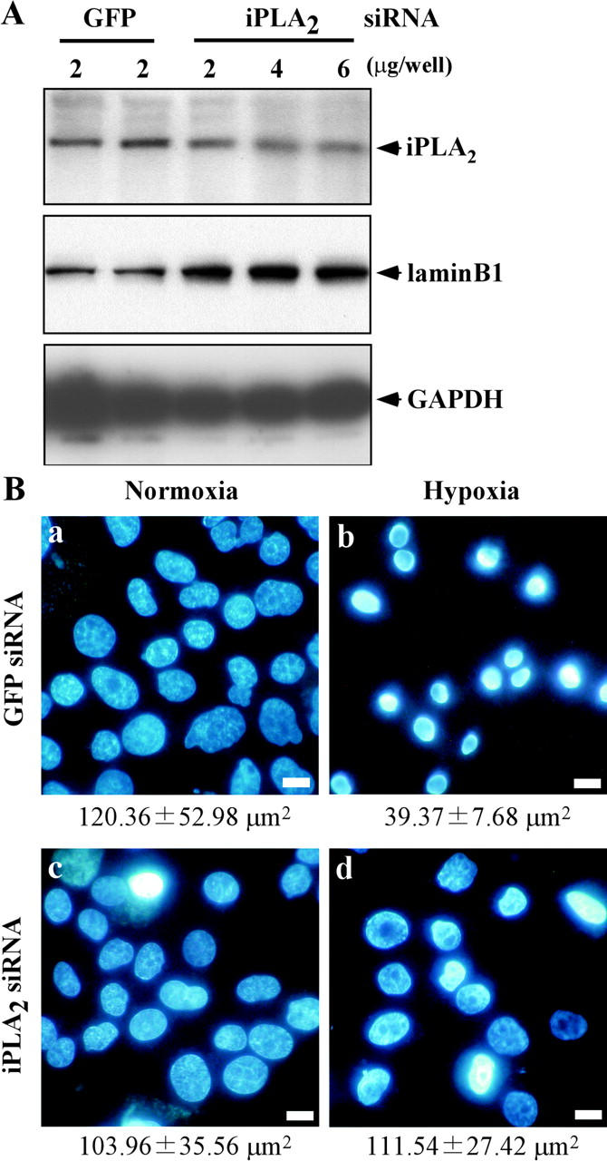
A reduced iPLA2 level inhibits hypoxic nuclear shrinkage. The indicated dose of siRNA for GFP or iPLA2 was transfected into CHO-k1 cells three times as described in Materials and methods. GFP was used as the negative control. (A) Expression of iPLA2 protein was assessed by Western blotting. The lysates from GFP siRNA- or iPLA2 siRNA-treated CHO-k1 cells were subjected to 15% SDS-PAGE followed by Western blotting with an antibody for iPLA2, lamin B1, or GAPDH (Lamin B1 and GAPDH were loading controls). (B) CHO-k1 cells were transfected with siRNA for GFP (2 μg/well) or siRNA for iPLA2 (6 μg/well), and then were exposed to normoxia or hypoxia for 36 h. Subsequently, the cells were stained with Hoechst 33342 and visualized under a fluorescence microscope. Bars, 10 μm. Note that siRNA for iPLA2 (2 μg/well) showed similar results (not depicted). The average nuclear area is shown under the photograph.