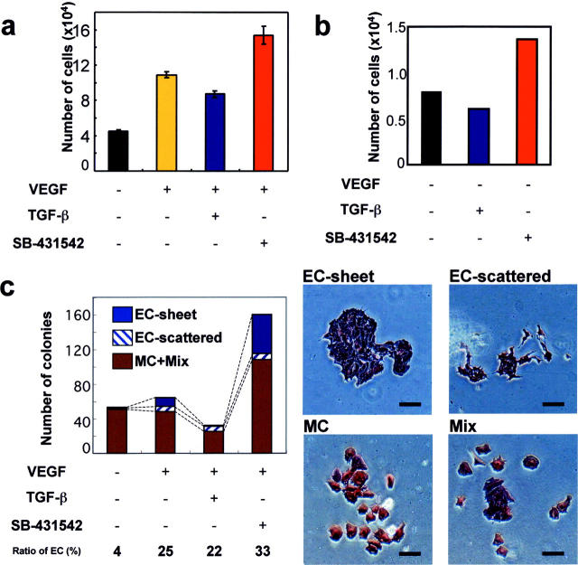
Figure 4.
Quantitative analyses of the effects of TGF-β signals on ESC-derived vascular differentiation. (a and b) Effect of TGF-β signals on proliferation of Flk1+ cells during vascular differentiation. 105 Flk1+ cells derived from CCE cells were cultured in the presence (a) or absence (b) of VEGF in combination with TGF-β or SB-431542, followed by determination of cell number after 3 d. Error bars represent SD. (c) Quantitation of colony formation, endothelial and mural cell production and endothelial sheet formation. Flk1+ cells were cultured sparsely with 10% FCS in the absence or presence of VEGF in combination with TGF-β or SB-431542 for 4 d, and stained for PECAM1 and SMA. Number of colonies per well was counted to determine the effect of TGF-β signals on colony formation of Flk1+ cells. Experiments were repeated at least three times with essentially same results. Four colony types were observed; pure endothelial cells forming sheet structures (EC-sheet): pure scattered endothelial cells (EC-scattered); pure mural cells (MC); and mixed colonies consisting of endothelial and mural cells (Mix). Bars, 50 μm.

