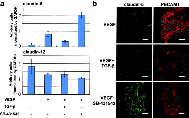
Figure 6.
Effect of TGF-β signals on expression of claudin-5 in ESC-derived endothelial cells. (a) Levels of expression of claudin-5 and -12 in vascular cells derived from MGZ5 cells cultured in the absence or presence of VEGF in combination with TGF-β or SB-431542 analyzed by quantitative real-time RT-PCR. Error bars represent SD. (b) Claudin-5 (left, green) and PECAM1 (right, red) double immunofluorescence staining of differentiated Flk1+ cells with 10% FCS and VEGF alone (top), or in combination with TGF-β (middle) or SB-431542 (bottom). To obtain similar numbers of endothelial cells, different numbers of Flk1+ cells were plated depending on the culture conditions. Bars, 200 μm.

