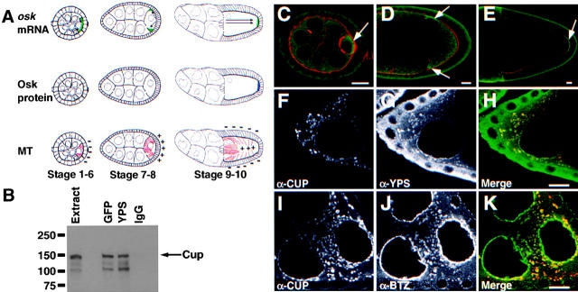
Figure 1.
Cup is a component of the oskar RNP complex. (A) Diagram showing the stage specific movements of oskar mRNA and the corresponding changes in Oskar translation and microtubule polarity. oskar mRNA, green; Oskar protein, blue; microtubules (MT), red. (B) Immunoblot for Cup (arrow) of immunoprecipitates from GFP-Exu extract using α-GFP (GFP), α-Yps (YPS), or rabbit IgG (IgG) antibodies. (C) Cup protein (green; arrows) is concentrated in at the posterior of the developing oocyte in stages 1–6 (stage 5 is shown; actin is in red). (D) Cup transiently accumulates at the anterior of the oocyte during stages 7 and 8 (stage 7 is shown). (E) Cup then accumulates at the posterior pole during stages 9 and 10 (stage 9 is shown). (F–H) Cup and Yps colocalize in cytoplasmic particles in nurse cells from stage 8 egg chambers. (F) α-Cup staining. (G) α-Yps staining. (H) Merged image. Cup is in red and Yps is in green. (I–K) Cup and Btz colocalize in cytoplasmic particles in nurse cells from stage 8 egg chambers. (I) α-Cup staining. (J) α-Btz staining. (H) Merged image. Cup is in red and Btz is in green. Bars, 10 μm.

