
Figure 2.
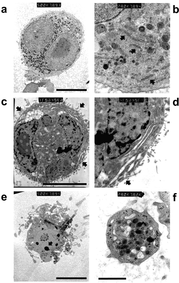
Mature MKs exhibit nuclear condensation typical of apoptosis, producing bodies indistinguishable from blood platelets. Purified mature MEG-01 MKs where examined by TEM. (a) MKs, shown at higher magnification in b to demonstrate a typical distribution of α-granules and demarcation membranes (arrows), contain nuclei with evenly dispersed heterochromatin. (c) MK, shown at higher magnification in d, exhibiting early cytoplasmic rearrangements consistent with platelet formation (arrows), with a nucleus displaying heterochromatin condensation typical of early apoptosis. (e) MK-bearing proplatelets show extensive condensation and fragmentation of the nuclear material (arrow). (f) TEM of MEG-01 MK culture supernatants reveals platelets with morphology consistent with that typically observed for blood platelets. Bars: 20 μm (a–e) and 1 μm (f).