
Figure 5.
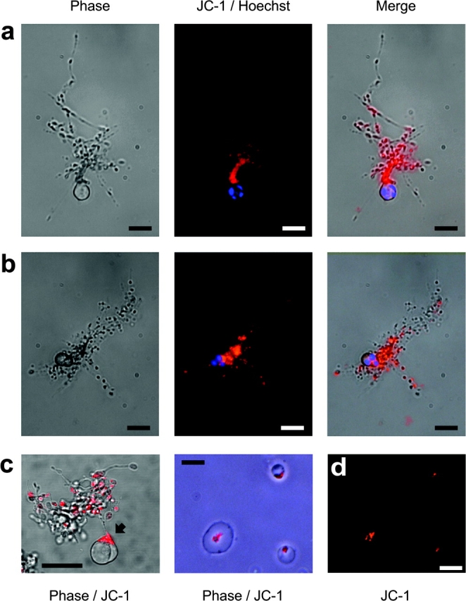
Functional platelet production is associated with the maintenance of ΔψM. (a and b) Mature MEG-01 MKs were double stained with the ΔψM-sensitive dye JC-1 (orange), and the nuclear staining vital dye Hoechst 33342 (blue). Mitochondria with an intact ΔψM are seen localized within platelet-sized nodes along extended proplatelets, whereas the cell bodies display nuclear condensation. (c) An MK stained with JC-1 only and analyzed by confocal microscopy reveals functional mitochondria with an intact ΔψM to be polarized within the cell body toward the remaining attached proplatelet (arrow). Bars in a–c, 20 μm. (d) Culture-derived platelets were also stained with JC-1 and allowed to adhere and spread on glass. Again, mitochondria with an intact ΔψM are seen coalesced within the body of the spreading platelet. Bar in d, 5 μm.