Figure 6.
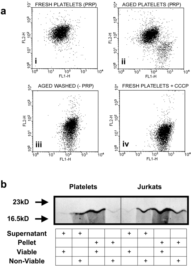
Senescent platelets show loss of ΔψM and release of mitochondrial cytochrome c . (a) Fresh PRP, aged PRP, or aged washed platelets, as indicated, were stained with the ΔψM-sensitive dye JC-1. (i) Flow cytometric analysis of fresh platelets display a typical fluorescent profile of high orange fluorescence, indicative of an intact ΔψM. (ii) Platelets aged in the presence of plasma survival factors show a small (∼10%) distinct subpopulation characteristic of a collapsed ΔψM, i.e., loss of FL2-H fluorescence with concomitant increase in FL1-H. (iii) By contrast, aged washed platelets display ∼100% loss of ΔψM. (iv) Treatment of fresh platelets with the protonophore mCCCP resulted in a collapse of the ΔψM, equivalent to that seen with aged platelets. (b) Aged platelets release mitochondrial cytochrome c into the cytoplasm. Fresh (viable) and aged washed (nonviable) platelets, as indicated, were lysed and cells were fractionated by centrifugation to yield a cytosolic supernatant and a subcellular pellet. Both supernatant and solubilized pellet were precleared with a control IgG before immunoprecipitating cytochrome c. Cytochrome c was detected by Western blot analysis, in which Jurkats treated with cycloheximide are provided as a positive control.

