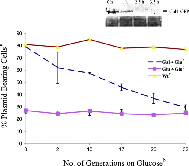
Figure 3.
Frequency of centromere switch in vivo. pDLB2064 was transformed into GAL1–UBI–CHL4 and wild-type (CDV39) cells and selected on Gal-uracil or Glu-uracil media. a,bCells were grown overnight and shifted to cGlu-ura (selective) media, and mitotic stability was determined at the generation times indicated. Colonies that were red or red and white sectored were scored as plasmid-bearing cells and are expressed as a percent of the total number of colonies plated. n = 3 transformants analyzed for Gal→Glu, 2 transformants for Glu→Glu, and 1 transformant for wild-type cells on glucose. Inset is a Western blot showing stability of the Chl4–GFP fusion protein in the degron strain. Cells from the degron strain were grown in galactose (0 h), and protein samples were prepared. Cells were washed and switched to glucose media to initiate proteolysis of the fusion protein. Protein samples were prepared at the time points indicated. The fusion protein was identified with anti-GFP antibodies in a Western blot analysis. The lower panel of the inset shows a region of the blot visualized by Ponceau S stain, indicating equal protein loading.

