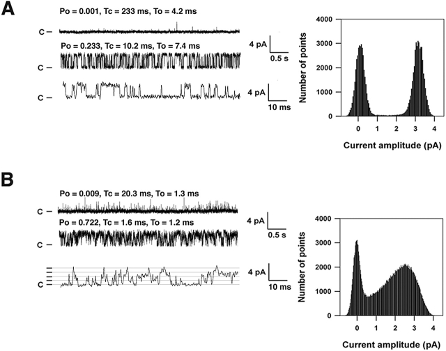
Figure 4.
Defective single channel properties of RyR1 isolated from skeletal muscle during HF. (A) Single channel traces from an RyR1 channel from normal canine skeletal muscle and corresponding current amplitude histogram. (B) Single channel traces from an RyR1 channel from HF canine skeletal muscle and corresponding current amplitude histogram. In A and B, upper tracings were recorded at 100 nM [Ca2+]cis, and bottom tracings were recorded after activation of the RyR1 channels with 1 mM ATP to increase Po. Open (To) and closed (Tc) dwell times and Po are shown for each condition above the representative tracings. In A and B, the bottom tracing is an expanded time scale. Lines indicating the current levels (0, 1, 2, 3, 4 pA) for the subconductance states of the PKA-hyperphosphorylated channel are shown in the bottom tracing in B. All point amplitude histograms for each channel are shown. Recordings were at 0 mV, the closed state of the channels is indicated as C, and the channel openings are upward deflections.

