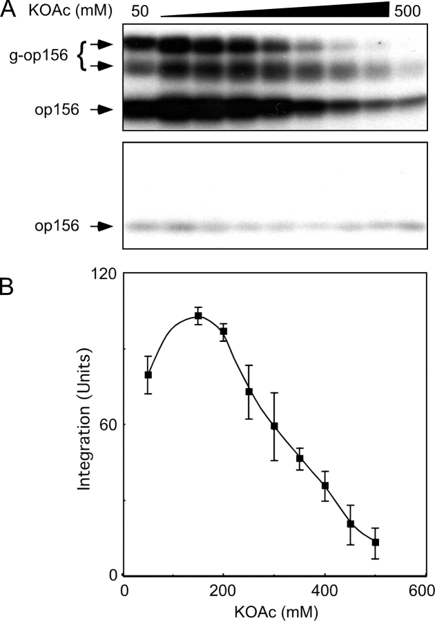
Figure 1.
Inhibition of translocation activity by high ionic strength. (A) SRP–ribosome–op156 complexes were adjusted to final KOAc concentrations ranging between 50 and 500 mM and incubated for 40 min at 25°C with 1.2 eq (as defined in Walter et al., 1981) of PK-RM (top) or 1.2 eq of protease-digested PK-RM (C1PK-RM) that lack intact SRα (bottom). After membrane-integrated forms of op156 were separated from nonintegrated op156 on alkaline sucrose gradients, the membrane pellets were analyzed by PAGE in SDS to resolve glycosylated (g-op156) and nonglycosylated (op156) polypeptides. (B) Membrane-integrated g-op156 was quantified with a phosphorimager and is expressed in units, where 100 U corresponds to the average of the 150 mM and 200 mM KOAc data points. The average and standard deviation were calculated using data from six experiments, one of which is shown in A.

