
Figure 1.
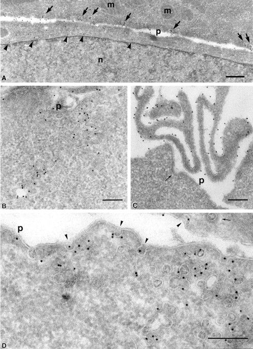
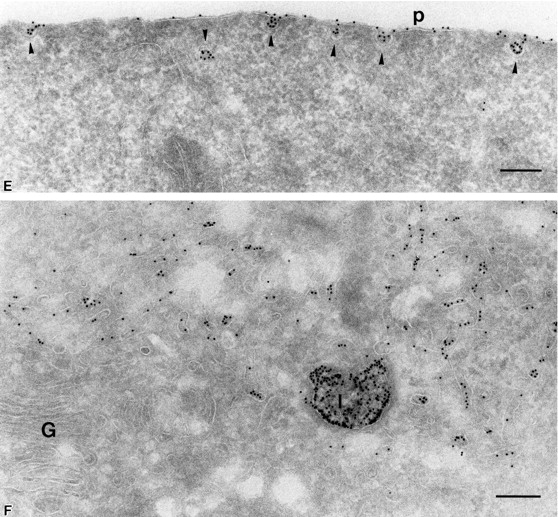
At steady state, PrP C is enriched in lamellae and caveolae both at the plasma membrane and inside the cell. (A–C) PrPC was immunogold labeled on ultrathin cryosections of CHO/30C3 cells (steady-state distribution of PrPC) using the R1 recombinant antibody Fab fragment against PrPC. Labeling was observed on the ER (A, arrowheads), plasma membrane caveolae-like structures (A, arrows), caveolae-like structures that appeared as flask-shaped invaginations on the plasma membrane (p) and interconnecting chains of caveolae-like structures deeper into the cytoplasm (B), and microvilli at the leading edge of the cell (C). Clathrin-coated pits (C, thin arrow) did not label. (D and E) Caveolin-1 was immunogold labeled on ultrathin cryosections of CHO/30C3 cells using anti-caveolin-1–gold labeling. (D) Caveolae-like structures and interconnecting chains of caveolae-like structures deeper into the cytoplasm labeled with a caveolin-1 antibody under steady-state conditions. (E) Protein A–gold uptake in an SHaPrPC-expressing cell, after a 10-min pulse. Gold labeling was observed on the plasma membrane and was enriched in caveolae (arrowheads). (F) After a 10-min pulse and 50-min chase, gold labeling was observed in small vesicles near the Golgi complex and lysosomes. G, Golgi complex; l, lysosomes; m, mitochondria; n, nucleus; p, plasma membrane. Bar, 200 nm (applies to all panels).