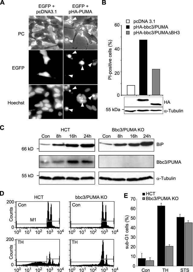
Figure 7.
Expression of bbc3/PUMA may be sufficient and required for ER stress–induced apoptosis in human cells. (A) Transient transfection of SH-SY5Y neuroblastoma cells with bbc3/PUMA leads to induction of apoptosis. Cells were cotransfected with expression plasmids encoding enhanced GFP (pEGFP) and either a hemagglutinin-tagged (HA) version of bbc3/PUMA (pHA-PUMA) or empty vector (pcDNA3.1). 12 h after transfection, cells were fixed and stained with Hoechst 33258. Arrowheads point to transfected cells with the apoptotic phenotype. Similar results were obtained in a separate experiment. (B) Quantitative FACS® analysis of PI uptake of EGFP-positive SH-SY5Y cells transiently cotransfected with expression vectors encoding hemagglutinin-tagged (HA) versions of Bbc3/PUMA, Bbc3/PUMA-ΔBH3, or empty vector (pcDNA3.1). Data were normalized to EGFP-only transfected controls and represent 10,000 events each. For evaluation of comparable transfection rates, whole-cell extracts of cells harvested 16 h after transfection were analyzed by Western blotting. Blots were probed with an anti-hemagglutinin or anti- α-tubulin antibody as loading control. (C) Protein levels of BiP, Bbc3/PUMA, and α-tubulin in human HCT116 control and Bbc3/PUMA-deficient cells treated with 1 μM thapsigargin or vehicle for the indicated time points. (D) FACS® analysis of sub-G1 cells of human HCT116 control and Bbc3/PUMA-deficient cells exposed to 1 μM thapsigargin (TH) or vehicle for 36 h. (E) Quantitative FACS® analysis of thapsigargin- and STS (3 μM)-induced apoptosis. Data from 10,000 events each are means ± SEM from n = 3 separate experiments per time point.

