
Figure 2.
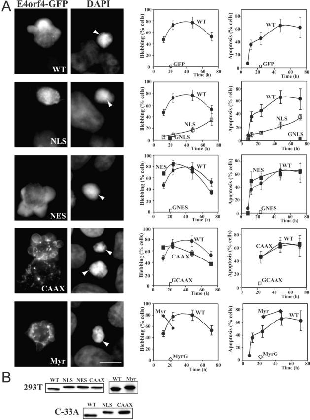
Functional activities of the E4orf4-GFP proteins. (A) 293T cells were transfected with the indicated Flag-E4orf4-GFP constructs (WT, NLS, NES, CAAX, and Myr), or with the relevant Flag-GFP controls (GFP, GNLS, GNES, GCAAX, and MyrG). At various times after transfection, the amounts of GFP-positive cells undergoing membrane blebbing were determined by fluorescence microscopy in live cells. Data are expressed as percent blebbing cells relative to the total number of GFP- positive cells. Other cultures of transfected cells were fixed and counterstained with DAPI and the nuclear morphology of GFP-positive cells was analyzed by fluorescence microscopy (representative phenotypes are shown). Apoptosis was determined by counting the number of cells expressing the relevant GFP proteins strictly in the targeted cell compartments that presented nuclear shrinkage and condensation (arrows). Data are expressed as the number of apoptotic cells relative to the total number of GFP-positive (means ± SE of at least four independent experiments, n > 2,000). Bars, 10 μm. (B) Aliquots of transfected cells were kept for Western blot analyses of expression levels using anti-Flag M2 antibody, or anti-GFP, 16–24 h after transfection.