
Figure 2.
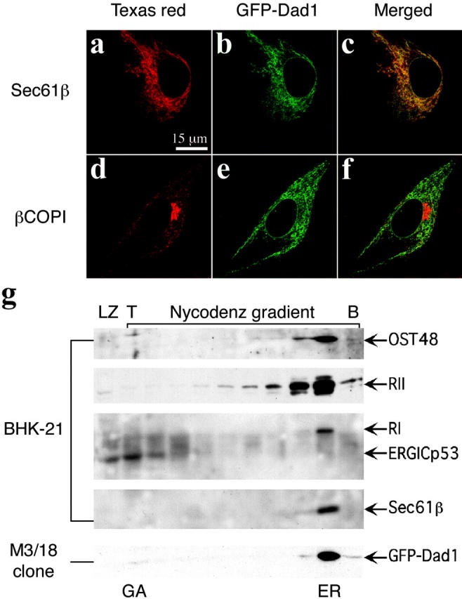
GFP–Dad1 expressed in clone M3/18 clone is located in the ER. M3/18 cell grown at 39.5°C were fixed in paraformaldehyde and permeabilized with Triton X-100. The cells were stained with primary pAbs against Sec61β (a) or βCOPI (d) and secondary antibodies were tagged with Texas red. Immunofluorescence micrographs were obtained with the LSM510 confocal microscope using the helium-neon laser. To localize GFP–Dad1 the argon-krypton laser was used (b and e). The merged images show that GFP–Dad1 is colocalized with Sec61β (c) and not with βCOPI (f). (g) GFP–Dad1 cofractionates with TC components. Postnuclear supernatants obtained from BHK-21 or M3/18 cells were fractionated on a Nycodenz gradient and equal aliquots were subjected to Western blot analysis using pAbs directed against the ER or ERGIC markers indicated next to the arrows marking the position of the respective proteins on the blots. The fractions containing Golgi and ERGIC or ER-derived membranes are indicated (GA or ER, respectively). GFP–Dad1 is only found in a fraction close to the bottom (B) of the gradient, which contains also other TC components. ERGICp53 is found exclusively in LZ and the two fractions next to the top (T) of the gradient.