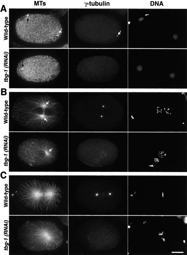
Figure 1.

The assembly of centrosomal asters in tbg-1(RNAi) embryos correlates with the presence of visibly condensed DNA. Embryos were fixed and stained for MTs (left), γ-tubulin (middle), and DNA (right). All panels are aligned with anterior to the left and posterior to the right. (A) During interphase, before visible DNA condensation, MTs grow from the still unseparated centrosomes in wild-type embryos (top, white arrows). Numerous cytoplasmic MTs are also present. In tbg-1(RNAi) embryos (bottom), cytoplasmic MTs are not affected but no centrosomal asters are detected. Remnants of the spindle from the second meiotic division of the oocyte pronucleus are visible in the anterior of both embryos (black arrows). (B) During mitotic prophase, the centrosomal asters in wild-type embryos have increased in size and are located between the two pronuclei (top, white arrows). In tbg-1(RNAi) embryos, MT asters form around unseparated centrosomes (bottom, arrow) positioned behind the sperm pronucleus. (C) After NEBD, a bipolar spindle is assembled in wild-type embryos (top). In tbg-1(RNAi) embryos, centrosomal asters have increased in size, but a mitotic spindle does not assemble (bottom). The DNA from the oocyte and sperm pronuclei remains in separate masses and does not align on a metaphase plate. Note that in some cases, our γ-tubulin antibody weakly stains condensed chromosomes (C, top middle). This staining does not disappear in tbg-1(RNAi) embryos (B, bottom middle). Bar, 10 μm.
