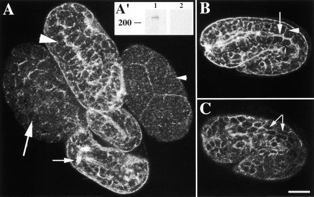
Figure 2.
α spectrin localizes to the cell membrane of most cells during embryogenesis. (A) Four representative wild-type embryos at different developmental stages labeled with AS1 and examined by confocal immunofluorescence microscopy. The small arrowhead indicates a four-cell stage embryo where a spectrin is localized to the cell junctions. An ∼100-cell stage embryo is shown with the large arrow; note cell membrane immunofluorescence. The large arrowhead indicates intestinal localization of a spectrin in a comma stage embryo. α spectrin localization in the nervous system of a threefold embryo is indicated by the arrow. (A') Western blot analysis of a mixed population of wild-type animals. Lane 1 indicates a ∼240-kD band recognized by AS1 on wild-type worm extracts. (Lane 2) Preincubation of AS1 with the GST–α spectrin fusion protein shows no reactivity to wild-type worm extracts. (B) A representative 1.5-fold embryo labeled with AS1. Basolateral and apical localization of a spectrin is shown. Arrow indicates apical region, and the arrowhead indicates basolateral region of the intestine. (C) The body wall muscle cells (identified by counter staining with myosin antibody [unpublished data]) of a representative 1.5-fold embryo are outlined by a spectrin (arrows). Bar, 10 μM.

