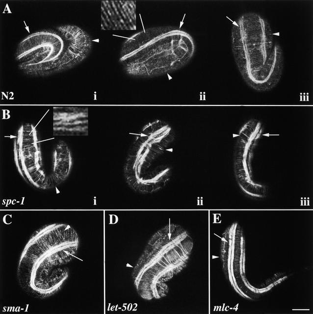
Figure 5.
α spectrin is required for the organization of the hypodermal apical actin cytoskeleton. Representative wild-type (A), spc-1(ra409) (B), sma-1(ru18) (C), let-502(h738) (D), and mlc-4(or253) (E) embryos were stained with FITC-phalloidin and examined by confocal fluorescence microscopy to visualize the circumferentially oriented actin cytoskeleton in the hypodermis. Arrowheads indicate the circumferential actin fibers, and arrows indicate the thin filaments of the body wall muscle. In wild type (A), the actin filaments are organized in a circumferential pattern in parallel bundles (arrowheads) that run perpendicular to the body wall muscle quadrant (arrows). Ai and Aii are threefold embryos (same developmental age as embryos shown in B–E, and Aiii is a 1.5-fold embryo. The inset shows parallel organization of the actin fibers. In spc-1 (α spectrin) embryos (B), several defects are observed in the highly regular pattern of the hypodermal actin cytoskeleton. Note the gaps (Bi and Biii, arrowheads), discontinuities, and clumping (Bii, arrowhead). Additionally, the thin filaments of spc-1 mutant embryos are abnormally oriented, and the body wall muscle quadrants are wider than the muscle quadrants in wild-type animals (Bii and Aii, arrows). The inset highlights the disorganization of the actin fibers. (C) Although slightly less severe, similar defects are observed in sma-1(βH spectrin) embryos. The arrowhead indicates a gap in the highly organized actin cytoskeleton. The hypodermal actin cytoskeleton in both let-502 (D) and mlc-4 (E) mutant embryos appears mostly wild type. In some instances, the spacing between some parallel actin bundles is abnormal in let-502(h738) embryos (see text). The same body wall muscle defect is observed in sma-1, let-502, and mlc-4 mutant embryos that is observed in spc-1 mutant embryos. All of these mutants have abnormally oriented thin filaments, and the body wall muscle is wider than normal (C and D, arrows). Bar, 10 μM.

