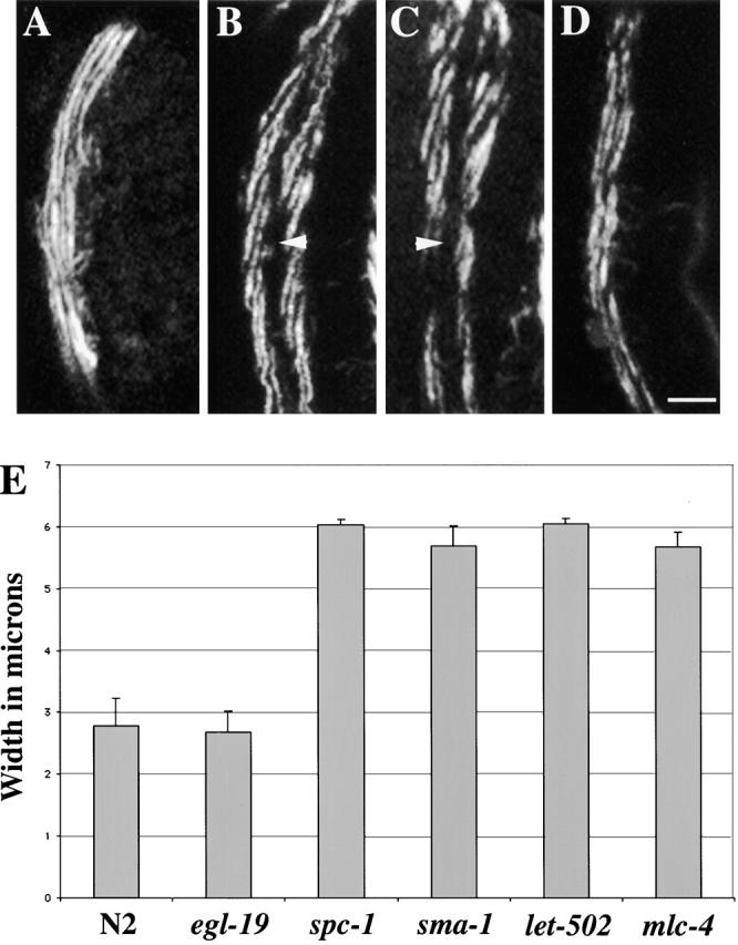
Figure 6.

Myosin filament organization is abnormal in elongation-defective mutants. Representative wild-type (A), spc-1(ra409) (B), sma-1(ru18) (C) , and egl-19(st556) (D) embryos were labeled with myosin antisera. The right dorsal muscle is shown in A–D, and anterior is up. In wild-type embryos (A), the myosin filaments are organized into a double row of A bands that runs approximately parallel with the longitudinal axis of the embryo. In both spc-1 (α spectrin) (B) and sma-1 (βH spectrin) (C) mutant embryos, the myosin filaments are organized into double rows of A bands, but the filaments are abnormally oriented. The myosin filaments in these mutants are almost oriented at a 20° angle to the longitudinal axis of the embryo. Additionally, the muscle quadrants of the α (B) and βH spectrin (C) mutant embryos are wider than normal (compare with A). Arrowheads in B and C indicate a large gap between the myofilament lattice in neighboring muscle cells. In egl-19 mutants (D), the body wall muscle quadrants and the myofilaments are normal (compare with A). Bar, 5 μM. (E) Width of the right dorsal muscle quadrant of N2 (n = 6), egl-19 (n = 5), spc-1 (n = 6), sma-1 (n = 5), let-502 (n = 6), and mlc-4 (n = 5) embryos. The width of a muscle quadrant in wild-type animals is ∼3 mm. Similarly, the muscle quadrants from the Pat mutant, egl-19, are ∼3-μM wide. The slow elongation mutants, spc-1, sma-1, let-502, and mlc-4, all have muscle quadrants that are ∼6-mm wide. Data is shown as the mean ± SD.
