
Figure 2. Overall structure of 13F6-1-2.
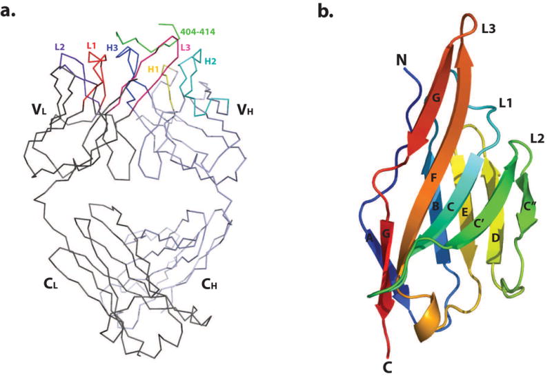
(a) Cα trace of the 13F6-1-2 Fab fragment complexed to the P404-P414 Ebola virus GP peptide. The structure is shown with bound GP peptide coloured in green, and the light and heavy chains in dark grey and light blue, respectively. CDRs L1, L2, L3, H1, H2, and H3 are coloured red, purple, pink, yellow, cyan, and blue, respectively. (b) Ribbon diagram of the 13F6-1-2 Vλx light chain variable domain. β-strands are labeled A, B, C, C′, C″, D, E, F, and G. This and subsequent figures were generated using MacPyMol 54.