
Figure 4. Antigen binding site.
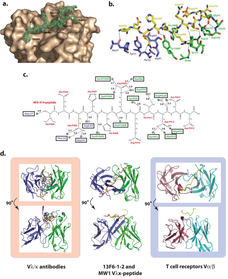
(a) Electron density for the P404-P414 GP peptide bound to 13F6-1-2. The initial difference σA-weighted Fo-Fc electron density map (green), contoured at 3σ superimposed onto the refined P404-P414 peptide and the molecular surface of 13F6-1-2. (b) Stick representation of the antigen binding site in which peptide residues are colored yellow, light chain residues are blue, and heavy chain residues are green. (c) 2-D schematic of the interactions between the peptide and Fab 13F6-1-2. Peptide resides are illustrated in black and labeled in red. (d) Comparison of general peptide binding orientations in Vλ/Vκ antibodies and Vα/β T cell receptors that bind peptides in a diagonal orientation. For all figures, the light and heavy chain and the TCR Vα and Vβ chains are coloured purple, green, red, and cyan, respectively. The light and heavy chains of all antibodies or α/β chains were superimposed. However for clarity, only the peptide is shown (PDB code 1CU4, red; 1TJG, brown; 1F58, blue; 1ACY, green; 1NAK, yellow; 1SM3, magenta; 1GGI, cyan; 1CFN, orange; and 1CE1, black). In the middle panel, the poly-Gln peptide bound to MW1 (PDB code: 2OTU), and GP peptide bound to 13F6-1-2 are shown in red and yellow, respectively.