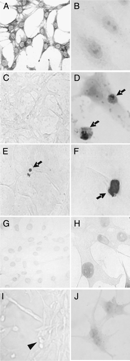
Figure 1.
Morphology of apoptotic MBA-15.4 osteoblasts. Healthy MBA-15.4 osteoblasts (A–C) were treated with dexamethasone (D–F and J) and assessed by immunocytochemistry for cleaved PARP (B and D) and by TUNEL (C, E–H). Normal morphology is shown using methylene blue stain (A). Apoptotic cells were identified on the basis of both positive staining and typical morphological changes of the cells (arrows; D–F). TUNEL staining was controlled using cover glasses pretreated with DNase to create breaks in the DNA of healthy cells (G and H), or as a negative control, highly apoptotic serum-starved cells with TUNEL but no terminal transferase enzyme (I). This produced a lighter stain than true apoptosis and no change in morphology (compare F with H), or no staining but apoptotic morphology with condensed, rounded cells (arrowhead; I). Secondary antibody only control (cPARP protocol) is shown for cells treated with dexamethasone (J).

