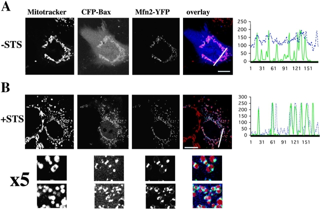
Figure 4.
Mfn2 protein forms mitochondria-associated punctate foci that colocalize with Bax during apoptosis. Cos-7 cells were cotransfected with CFP-Bax (blue) and Mfn2-YFP (green), incubated without (A) and with (B) 1 μM STS for 90 min, and analyzed by confocal microscopy after staining with Mitotracker CMXRos (red). In untreated cells (A) Bax is mostly cytosolic and Mfn2 is concentrated in punctate mitochondrial foci. In apoptotic cells (B) Bax localizes to foci containing Mfn2. The line scans plot the intensity of CFP-Bax (blue) and Mfn2-YFP (green). Bars, 20 μm.

