Figure 3.
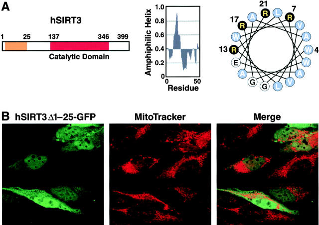
The NH 2 -terminal region of hSIRT3 is required for mitochondrial import. (A) Schematic diagram of hSIRT3. The orange box illustrates the region involved in mitochondrial targeting (left). Parts of the NH2-terminal region show a high probability of forming an amphiphatic α-helix (middle). A helical wheel plot of residues 4–21 reveals a cluster of basic amino acids (black) on one side of the putative helix (right). (B) HeLa cells grown on coverslips were transfected with hSIRT3Δ1–25–GFP for 36 h, stained with MitoTracker, and analyzed by confocal laser scanning microscopy. (left) GFP fluorescence emitted by the fusion protein (green). (Middle) MitoTracker signal (red). (Right) Merged image.

