
Figure 5.
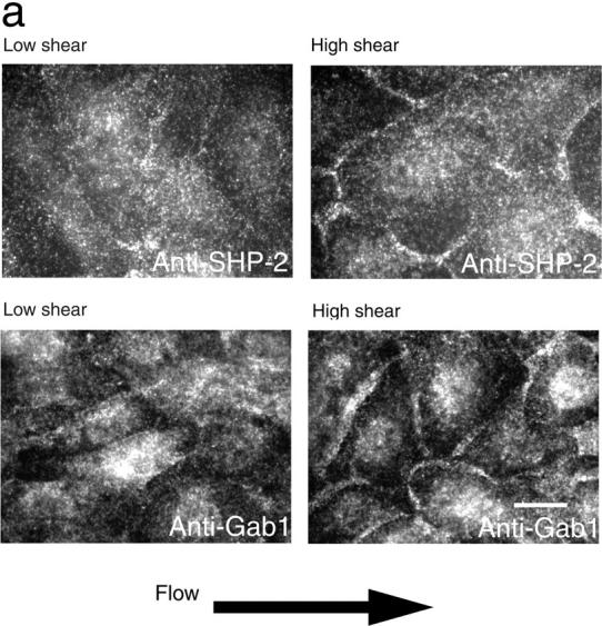
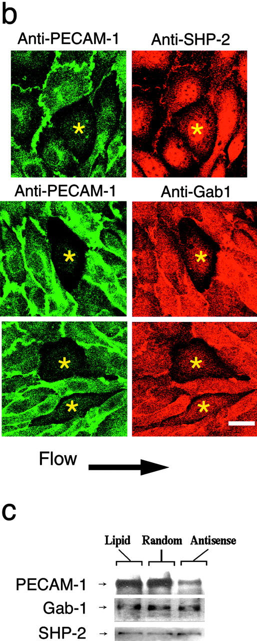
FSS-dependent localization of SHP-2 and Gab1 at the cell junction. (a) Immunofluorescence localization of endogenous SHP-2 and Gab1 in confluent BAECs exposed to 0.1 dyn/cm2 (low shear) or 15 dyn/cm2 (high shear) for 5 min. Both SHP-2 and Gab1 were distributed diffusely in ECs under low shear stress, but they translocated to the cell border in cells exposed to high shear stress. (b) BAECs were treated with PECAM-1 antisense S-oligo (low transfection condition) and allowed to form a monolayer. They were exposed to 15 dyn/cm2 of FSS for 5 min, fixed, stained doubly with anti–PECAM-1 and anti–SHP-2, or with anti–PECAM-1 and anti-Gab1, and observed using a confocal microscope. Although both SHP-2 and Gab1 accumulated at the cell junction in ECs expressing PECAM-1, they did not accumulate at the cell junction in cells with reduced PECAM-1 expression (asterisks). For the experiments shown in a and b, ECs were exposed to flow immediately after adding 1 mM Na3VO4 to the medium. Bars, 10 μm. (c) BAECs were treated with PECAM-1 antisense S-oligo (Antisense), scrambled sequence S-oligo (Random), or lipid carrier (Lipid) using the high transfection efficiency condition as in Fig. 2. Cell lysates were immunoblotted using anti–PECAM-1cyt, anti-Gab1, and anti–SHP-2. Although the antisense treatment greatly reduced PECAM-1 expression, Gab1 and SHP-2 expression levels were not affected. The same results were obtained in three separate experiments.