
Figure 7.
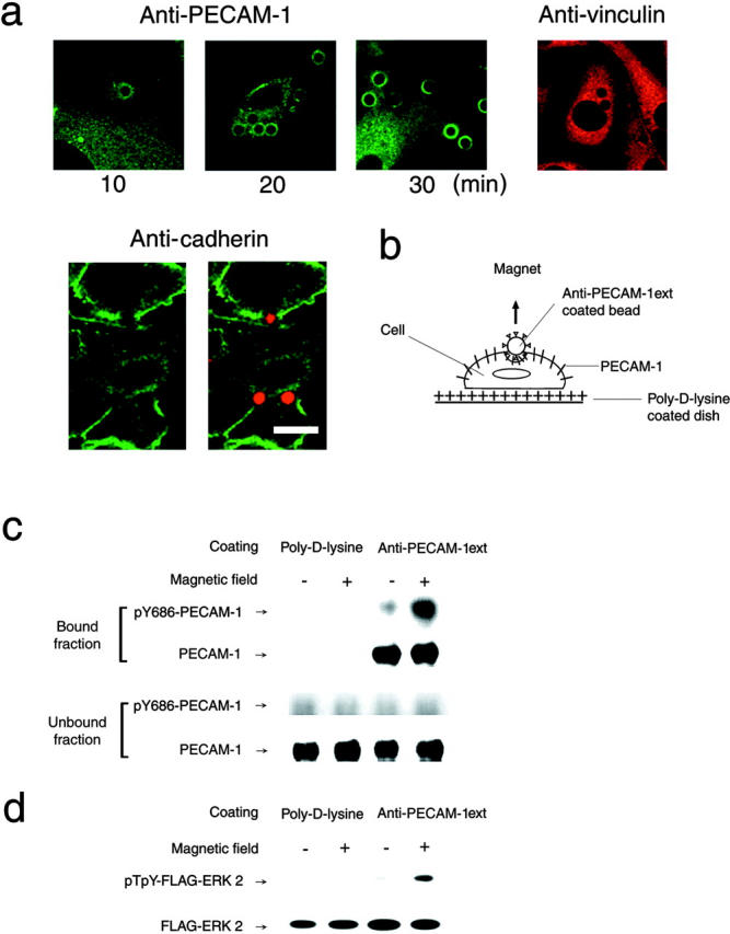
PECAM-1 tyrosine phosphorylation and ERK activation by direct application of mechanical force to PECAM-1. (a) Magnetic beads coated with rabbit anti–PECAM-1ext were allowed to attach to sparsely cultured BAECs for 10–30 min. Cells were fixed and stained with chicken anti–PECAM-1. Confocal micrographs show time- dependent PECAM-1 accumulation under the beads. To see if molecular aggregates similar to the focal adhesion or the adherens junction might be formed under the beads, cells were stained with antivinculin or anti–pan-cadherin. No accumulation of vinculin and cadherin was detected with the beads in 20 min. Beads with autofluorescence were used for anti–pan-cadherin staining. Anti–pan-cadherin staining (left) and a merged image (right) of the antibody (staining green) and autofluorescent beads (red) are shown. Note that anti–pan-cadherin stains the EC junction but not with the beads. Bar, 10 μm. (b) A schematic representation of the experiment. BAECs plated sparsely on a poly-l–coated surface were cultured for 1 h in serum-free DF medium. Magnetic beads coated with anti–PECAM-1ext or poly-l were allowed to adhere to the cell surface for 20 min and a strong magnet was put over the cell for 10 min. In real experiments, beads covered practically the entire apical surface of cells. (c) Cells were lysed and beads collected. Proteins bound to beads were eluted with SDS sample buffer (Bound fraction). The remaining cell lysates were mixed with an appropriate amount of SDS sample buffer (Unbound fraction). Both samples were immunoblotted by anti-686pY and reprobed with anti–PECAM-1. PECAM-1 bound to the beads was tyrosine phosphorylated in a magnetic field–dependent manner. For detail, see text. (d) BAECs were transiently transfected with FLAG–ERK2 and subjected to the same experiment as in c. From cell lysates, FLAG–ERK was immunoprecipitated by anti-FLAG and immunoblotted with anti–phospho-ERK and reprobed with anti-ERK. ERK activation was observed in cells whose PECAM-1 was tugged by the antibody-coated beads. The experiments illustrated in c and d were repeated five to eight times with the same results.