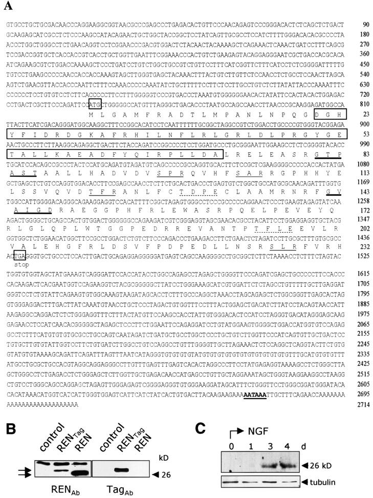
Figure 2.

Sequence and expressoin of REN cDNA and protein. (A) Nucleotide and predicted amino acid sequence of mouse REN cDNA. The first and last codons of the ORF are boxed and the putative polyadenylation signal is in bold-face type and underlined. Putative casein kinase 2 (dotted underline), protein kinase C (underline), and N-myristoylation (thick underline) sites and BTB/POZ motif (boxed) are shown. These sequence data are available from at EMBL/GenBank/DDBJ accession no. AF465352. (B) Western blot analysis of REN protein in cell lysates from COS7 mock-transfected cells (control), transiently transfected with pCXN2-REN-myc encoding myc epitope-tagged REN protein (RENTag) or with pCXN2-REN encoding wild- type REN (REN) (arrows). Immunoblotting was performed with either anti-REN (RENAb) polyclonal or anti-myc (TagAb) monoclonal antibodies. (C) Western blot analysis of REN protein in cell lysates from PC12 cells treated for the indicated times with NGF, revealed by anti-REN antibody. α-Tubulin staining is also shown, as a loading control.
