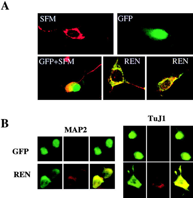
Figure 7.
Exogenous expression of REN enhances neuronal marker expression in ST14A and N2a neural progenitor cells. Confocal microscopy of combined coimmunostaining of MAP2 or TuJ1 (red) and either myc epitope (green) or GFP (green; overlapping signals are yellow). (A) Untransfected (SFM) or GFP-transfected (GFP + SFM) ST14A cells grown in SFM for 7 d display a differentiated morphology and positive staining for MAP2. Cells transfected with GFP encoding expression vector and cultured in growth medium for 7 d, display GFP immunofluorescence and no staining for MAP2 (GFP panel). Cells transfected with myc epitope-tagged REN protein encoding vector and cultured in growth medium for 7 d, display coexpression of both myc epitope and MAP2 immunofluorescence (REN panels). (B) N2a cells transfected with REN expression vector (REN) display coexpression of REN and either MAP2 or TuJ1, 3 d after transfection. In contrast, cells transfected with GFP encoding expression vector and cultured for 3 d, display GFP immunofluorescence and no staining for either MAP2 or TuJ1. The pictures shown are from a representative experiment (out of three) with MAP2 or TuJ1 expression scored from 150 to 300 transfected cells examined.

