
Figure 3.
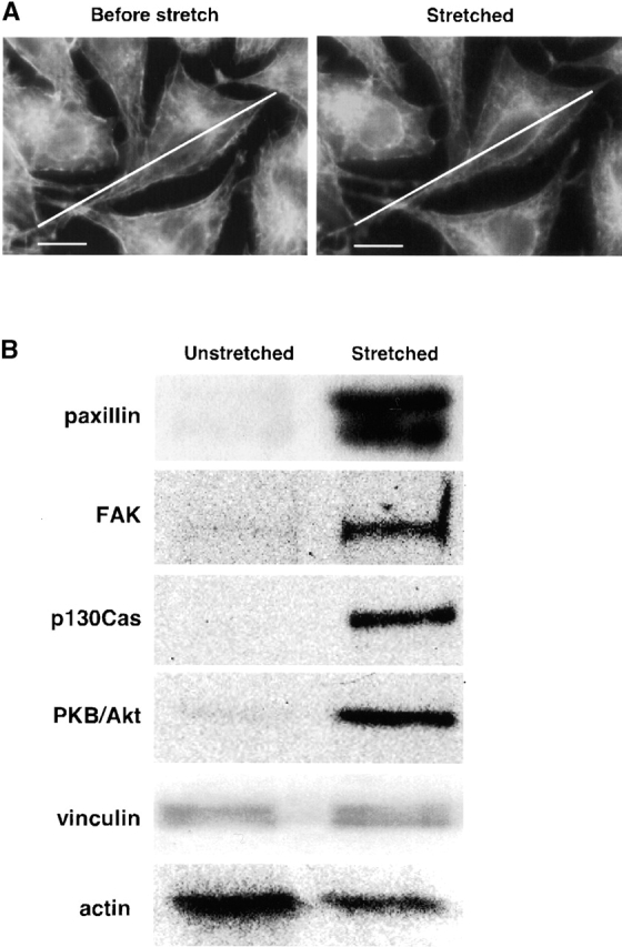
Focal contact proteins bind preferentially to stretched cytoskeletons. (A) Micrographs showing that Triton X-100–insoluble cytoskeletons are stretched 10%. Triton X-100–insoluble cytoskeletons on a collagen-coated silicone membrane (StageFlexer®; Flexcell International) were incubated with rhodamine-phalloidin (Molecular Probes) for 2 min, and washed three times with ISO (+) buffer. Images were obtained with an Olympus BX50 microscope with a 60×, 0.9 NA water immersion objective. Focus was adjusted to identify the peripheral margins (lower surface of cells), and images were obtained before stretch (Before stretch) and 5 min after stretch (Stretched). The diagonal lines show the length of the cell before stretch. (B) Western blots of focal contact proteins bound to unstretched and stretched cytoskeletons. L-929 cytoplasmic proteins tagged with a photocleavable biotin (NHS-PC-LC-biotin) were added to Triton X-100–insoluble cytoskeletons of L-929 cells on a stretchable silicone dish (Sawada et al., 2001), and cytoskeletons were stretched or left unstretched (Fig. 1). After washing, bound cytoplasmic proteins were eluted with 1 ml of 1 M NaCl in HYPO buffer (as described in Materials and methods), precipitated with avidin beads (immobilized neutravidin; Pierce Chemical Co.) after sevenfold dilution with HYPO buffer, and released from the bead complex by irradiation with 302 nm UV light (10 min). After photocleavage, proteins were eluted with 120 μl of HYPO buffer, and 40 μl of the sample was subjected to 10% SDS-PAGE followed by immunoblotting with antibodies to paxillin, FAK, p130Cas, PKB/Akt (Transduction Laboratories), vinculin (Upstate Biotechnology), or actin (Santa Cruz Biotechnology). Bar, 10 μm.