
Figure 4.
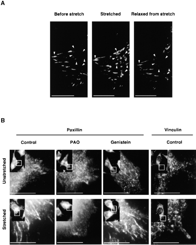
Fluorescence micrographs of the stretch-dependent distribution of GFP paxillin, endogenous paxillin, and vinculin in intact L-929 cells. (A) L-929 cells transiently transfected with GFP paxillin were cultured on collagen-coated silicone membranes in a StageFlexer system. Cells were stretched by 10%, held for 5 min, and subsequently relaxed by allowing the stretched silicone membrane to return to its original size. GFP fluorescence was observed with an Olympus BX50 microscope using a 60×, 0.9 NA water immersion objective before stretch (left), 2 min after stretch (middle), and 2 min after relaxation of stretch (right). Arrowheads indicate sites of GFP paxillin assembly dynamics: Before stretch, Stretched, and Relaxed from stretch. (B) Antipaxillin or antivinculin antibody distribution in L-929 cells cultured on silicone membranes was measured either for unstretched (top), stretched (for 2 min; bottom), stretched PAO treated (20 μM, for 10 min), or stretched genistein treated (100 μM, for 10 min) cells. Cells were fixed with 3.7% formaldehyde/PBS, permeabilized with 0.1% Triton X-100/PBS, and subjected to immunostaining using an antipaxillin or an antivinculin antibody. Stretched samples were relaxed after fixation. The rectangle in the inset (low magnification) indicates the area of each micrograph. Bars, 10 μm.