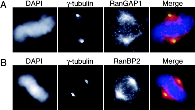
Figure 2.
RanGAP1 does not localize at the immediate vicinity of spindle poles. HeLa cells were stained with either anti-RanGAP1 or anti-RanBP2 and anti–γ-tubulin antibodies after permeabilization and fixation to observe the localization of these proteins at spindle poles. (A) γ-Tubulin (green) and RanGAP1 (red), as recognized by corresponding antibodies. (B) γ-Tubulin (green), RanBP2 (red), and DNA were visualized by staining with DAPI (blue).

