
Figure 2.
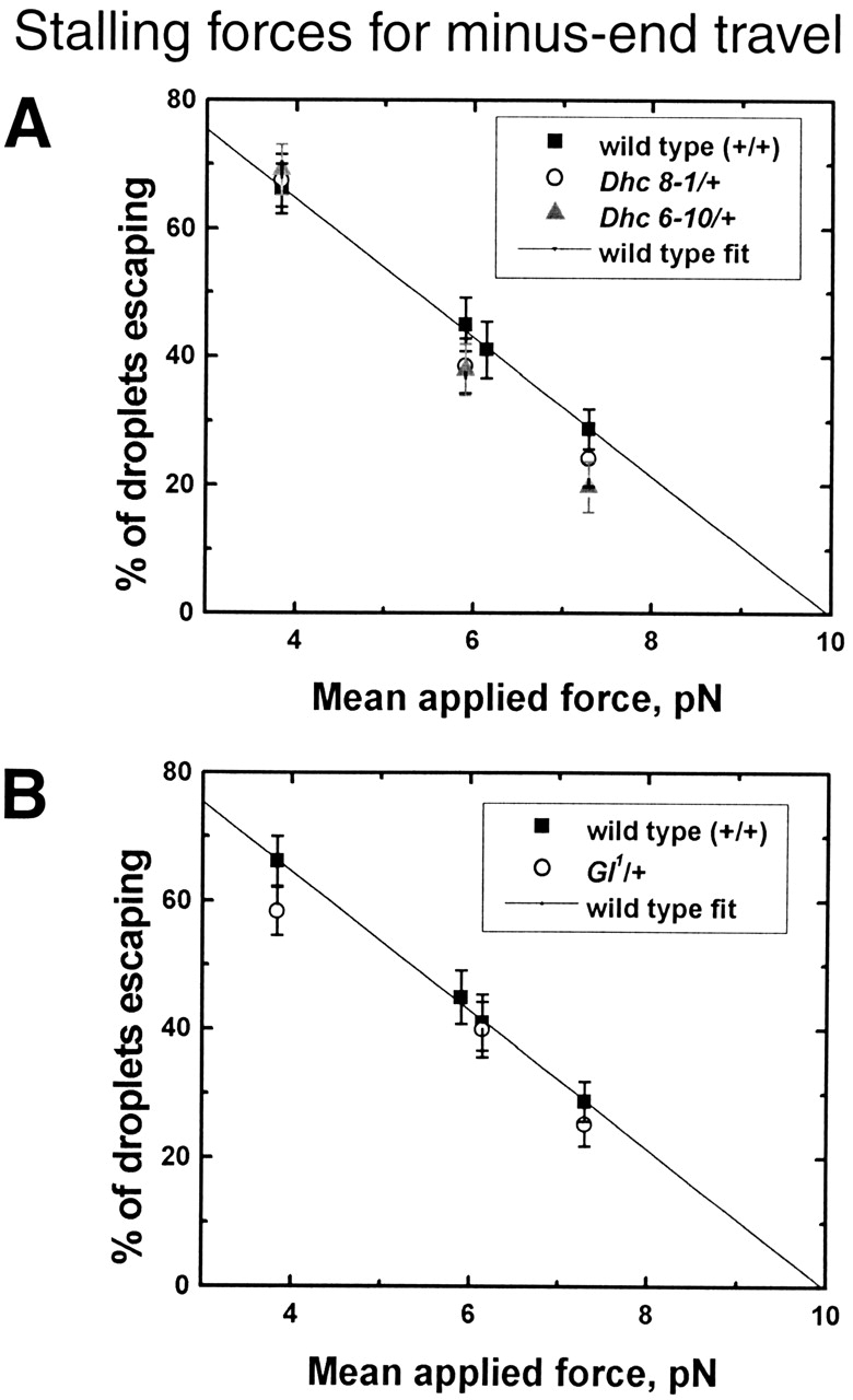
Droplet stalling forces for minus-end travel. The panels show the percentage of droplets stalled in different genetic backgrounds as a function of force applied by optical tweezers. (A) Wild- type versus Dhc64C 6–10/+ and Dhc64C 8–1/+; (B) Wild-type versus Gl1/+. To avoid bias, force measurements were performed in a blind fashion, with the genotype of the embryo being measured unknown to the person performing the force measurement. Each data point is derived from measurements on six to seven embryos in phase II, with ∼30 minus-moving droplets tested per embryo. See Materials and methods for a discussion how applied force and stalling force are related.