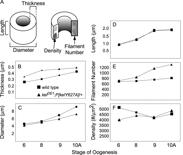
Figure 7.
Quantitation of thin-section electron micrograph data. Wild-type (▪) and kel DE1;P[kelY627A]/+ (▴) ring canals are compared over four stages of development (6, 8, 9, and 10A). At least seven ring canals were measured at each stage. (A) A ring canal can be characterized as a tube with three dimensions: length, diameter (to the outer rim), and thickness of the inner rim. Counting the total number of actin filaments per ring canal section leads to the filament number. The density of actin filaments was calculated by counting filaments in 10 nm2 segments of the inner rim that contained actin. (B) Beginning at stage 6 of oogenesis, there is a statistically significant increase (P < 0.01) in inner rim thickness in kel DE1;P[kelY627A]/+ compared with wild type. (C) Beginning at stage 9, kel DE1;P[kelY627A]/+ ring canal diameters are smaller than wild type. (D) The lengths of the outer rims are similar during all observed stages. (E) Actin filament number increases in kel DE1;P[kelY627A]/+, and by stage 10A there are nearly 50% more filaments per section compared with wild type. (F) Although there may be an initial difference in actin density, wild-type and kel DE1;P[kelY627A]/+ ring canals ultimately have similar actin densities (measurements of wild-type stages 8 and 10A are from Tilney et al., 1996). The size of each data point represents the standard error of measurement.

