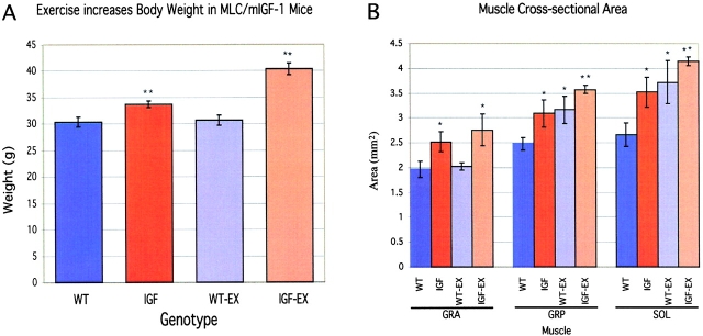
Figure 2.
Skeletal muscle hypertrophy of inactive and exercised WT and mIGF transgenic mice. (A) Comparison of body weights shows a significant increase in IGF and IGF-EX compared with WT animals, with IGF-EX animals weighing the most. (B) Muscle cross-sectional area increased significantly in gracilis anterior, gracilis posterior, and soleus muscles, indicating hypertrophy in these muscles. Since the MLC/mIGF-1 transgene is expressed at very low levels in soleus (Musarò et al., 2001) the increase in the average cross-sectional area of soleus muscle may reflect a secondary effect of increased loading in neighboring muscles, resulting in a transition to a faster fiber type.

