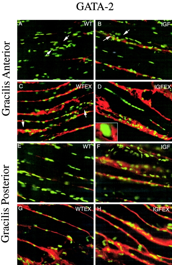
Figure 5.

GATA-2 localization on gracilis anterior or posterior muscles with Hoechst (green fluorescence) and AChE (brightfield) stains. (A–D) Gracilis Anterior with AChE precipitate between arrows. (A) WT, (B) MLC/mIGF-1 transgenic, (C) WT-EX, (D) MLC/mIGF-1 transgenic exercised, and (D, insert) GATA-2 is excluded from the nucleus. (E–H) Gracilis posterior. (E) WT, (F) MLC/mIGF-1 transgenic, (G) WT-EX, (H) MLC/mIGF-1 transgenic exercised. GATA-2 protein is upregulated in IGF and WT-EX, with a further increase in IGF-EX.
